A Purple Sapphire Containing Interesting Inclusions: Formation Insights
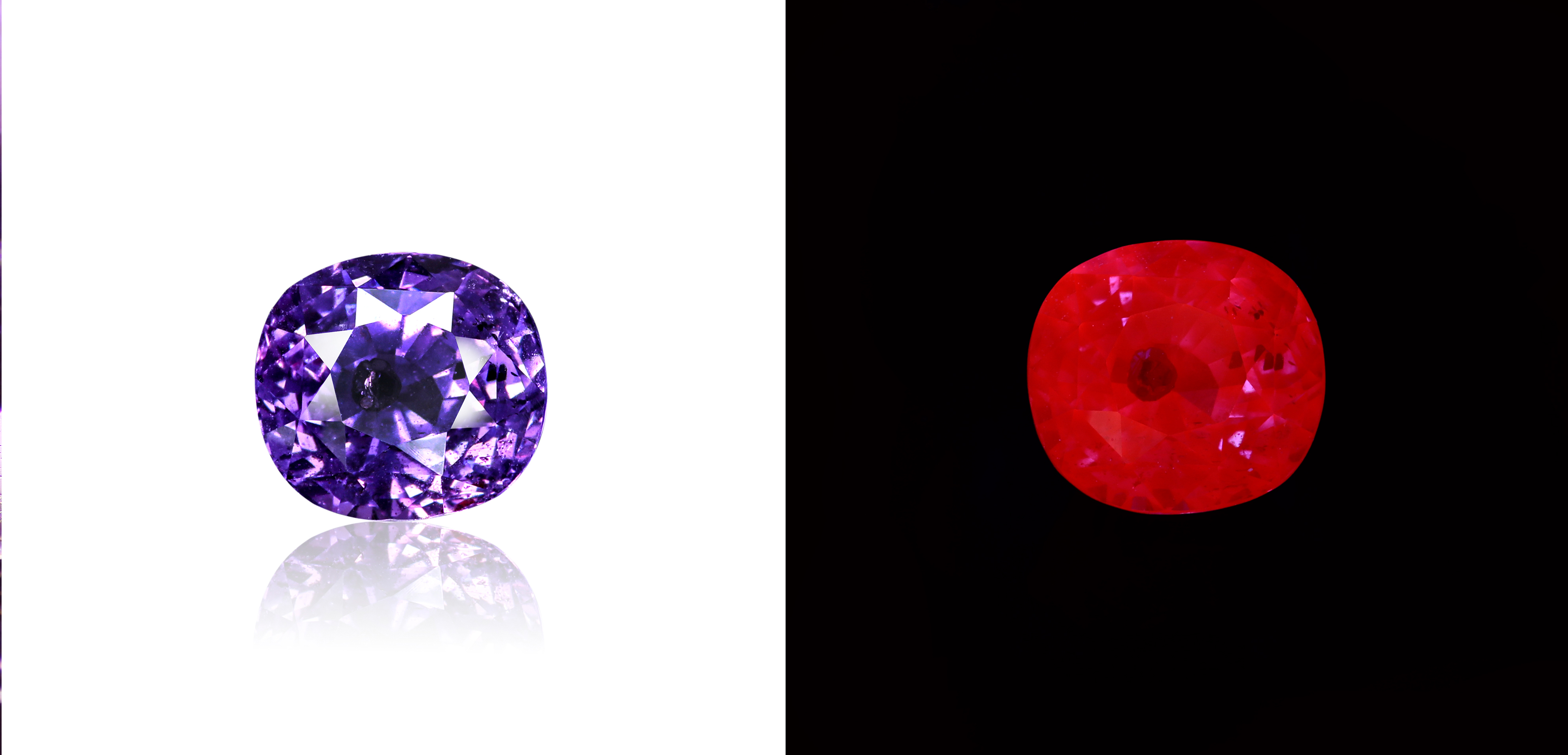
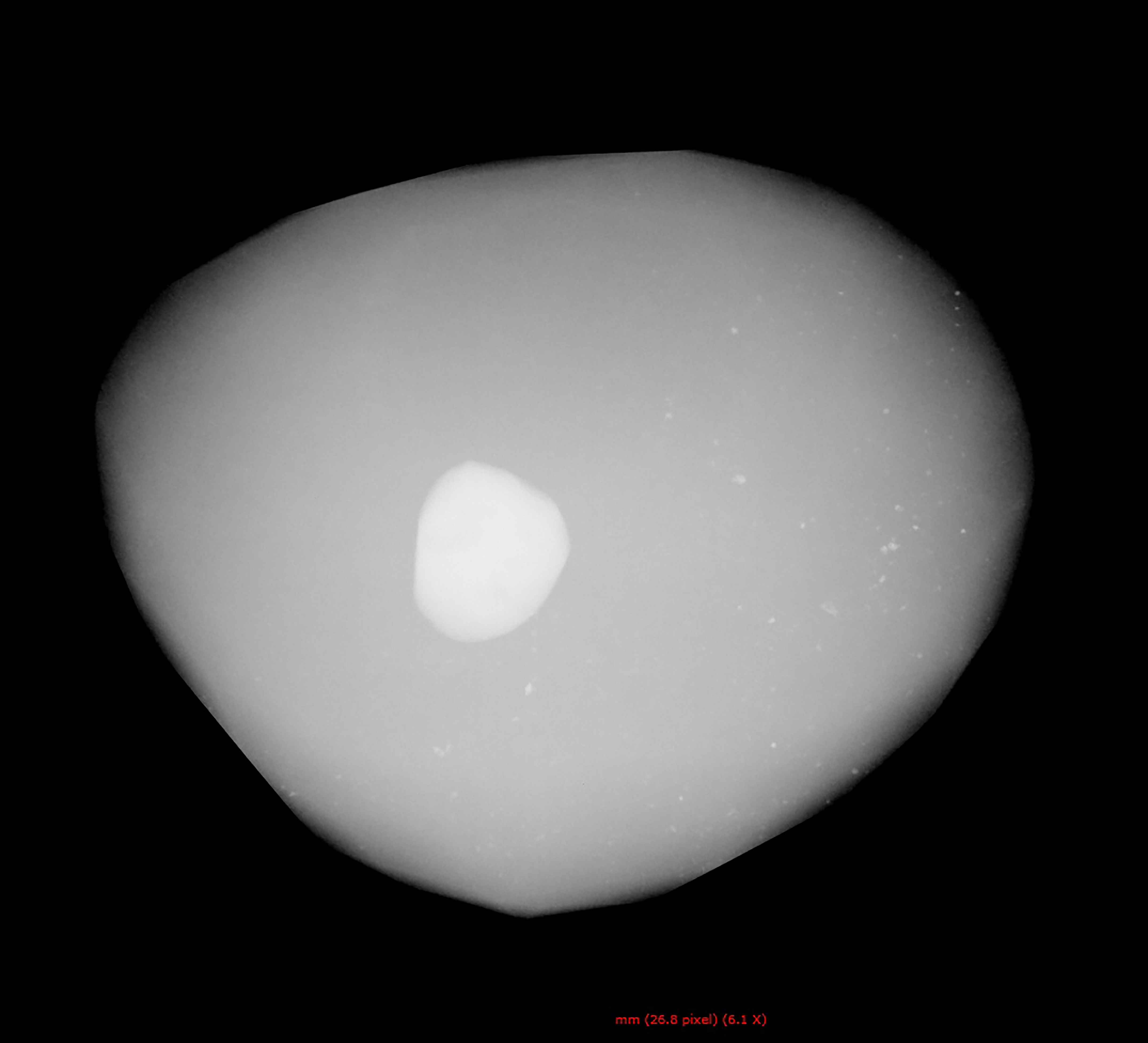
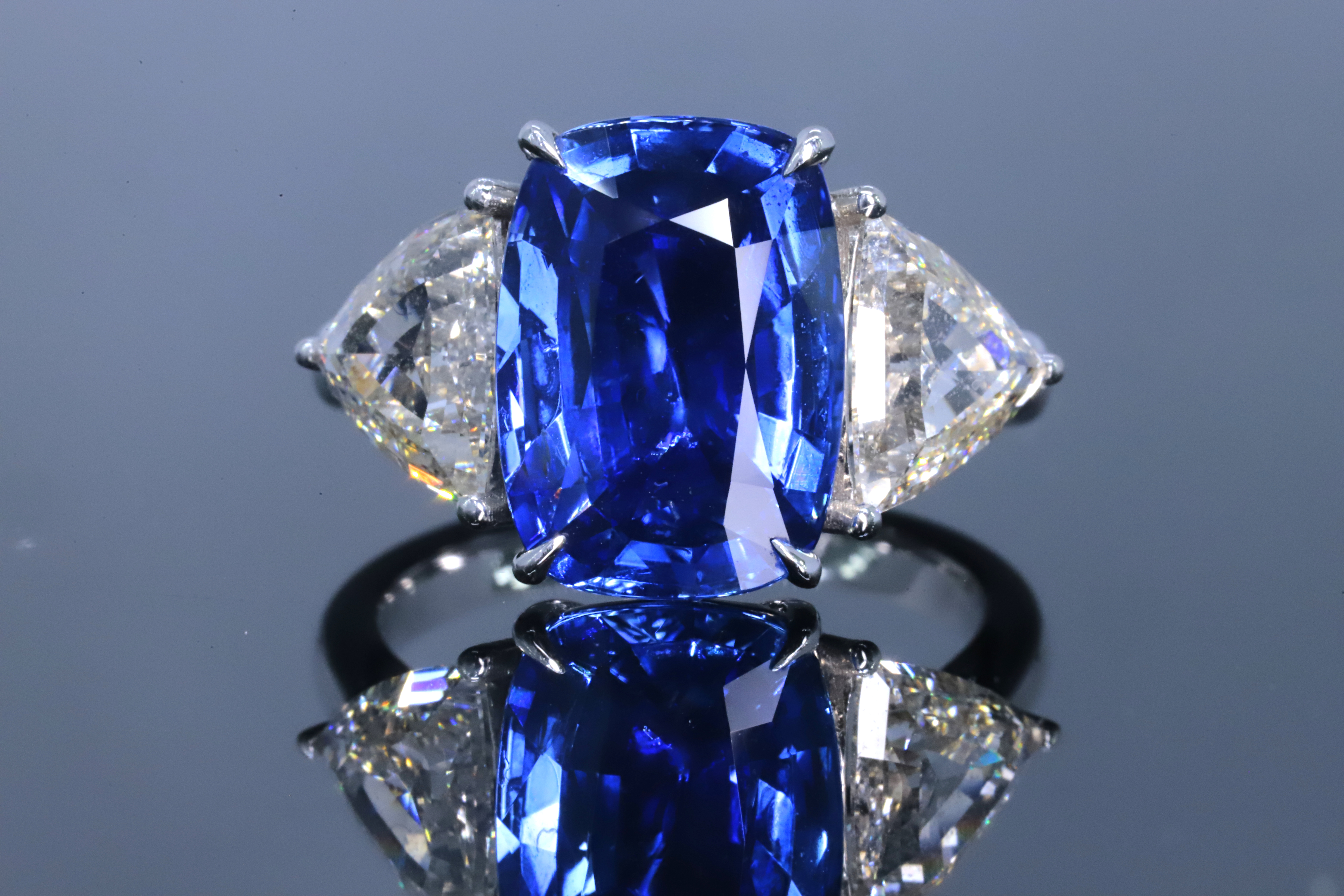
Figure 40: (a) A 3.56 ct purple sapphire was examined for this report. (b) It exhibits strong red fluorescence to long-wave UV radiation, except for inert areas corresponding to inclusions. (c) A large eye-visible inclusion under the table shows high density in this X-ray image. Photos (a, b) by Fei Hu and X-ray image (c) by Ziyun Zhang.
Recently, the authors examined a cushion-shaped purple sapphire weighing 3.56 ct (Figure 40a). The RIs were 1.762–1.770, yielding a birefringence of 0.008, consistent with sapphire. FTIR spectroscopy confirmed its identification as corundum. The stone displayed strong red fluorescence to long-wave (365 nm) UV radiation (Figure 40b). Chemical analysis by EDXRF spectroscopy revealed appreciable Cr (about 720 ppm) and Fe (approximately 1,200 ppm), along with some Ti (around 230 ppm).
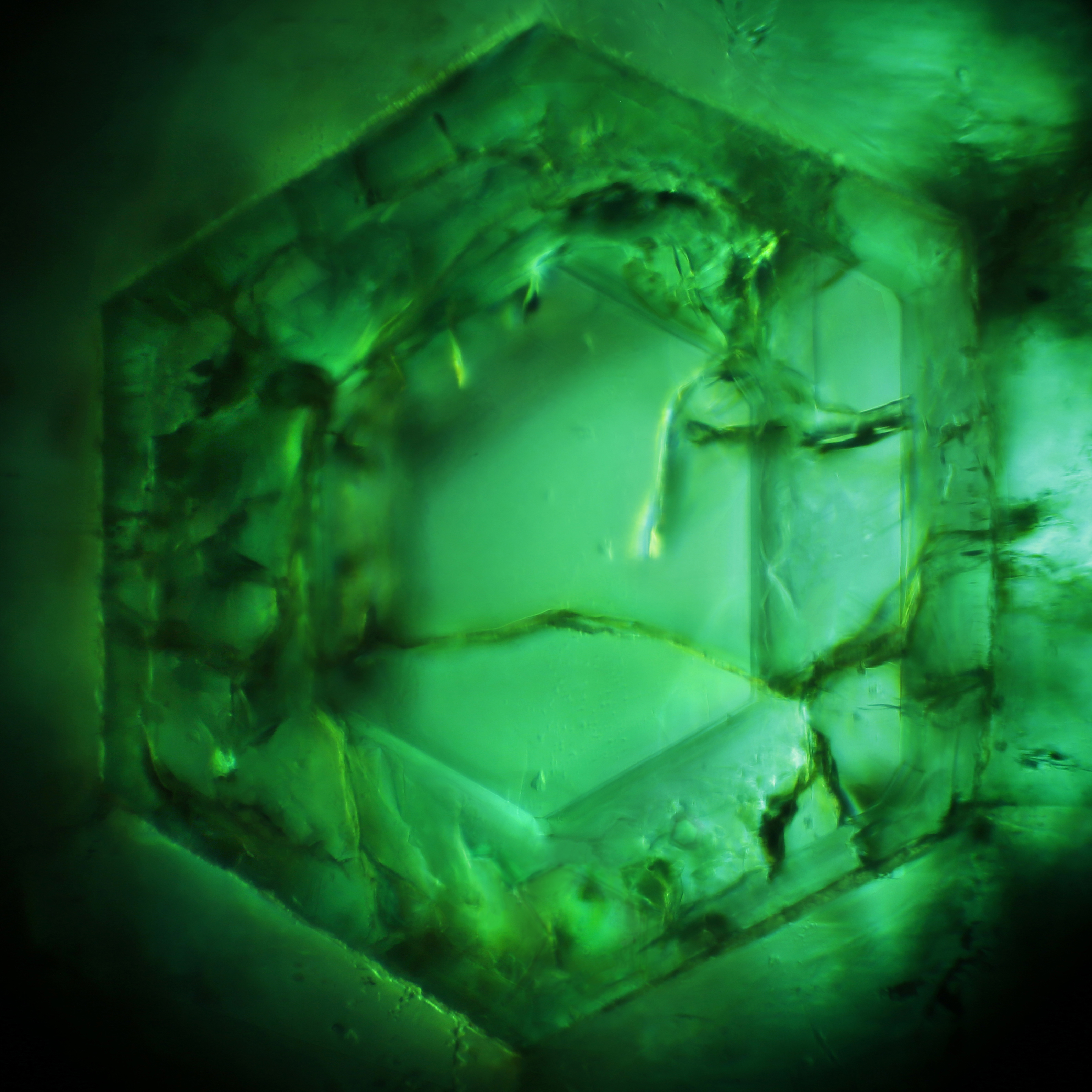
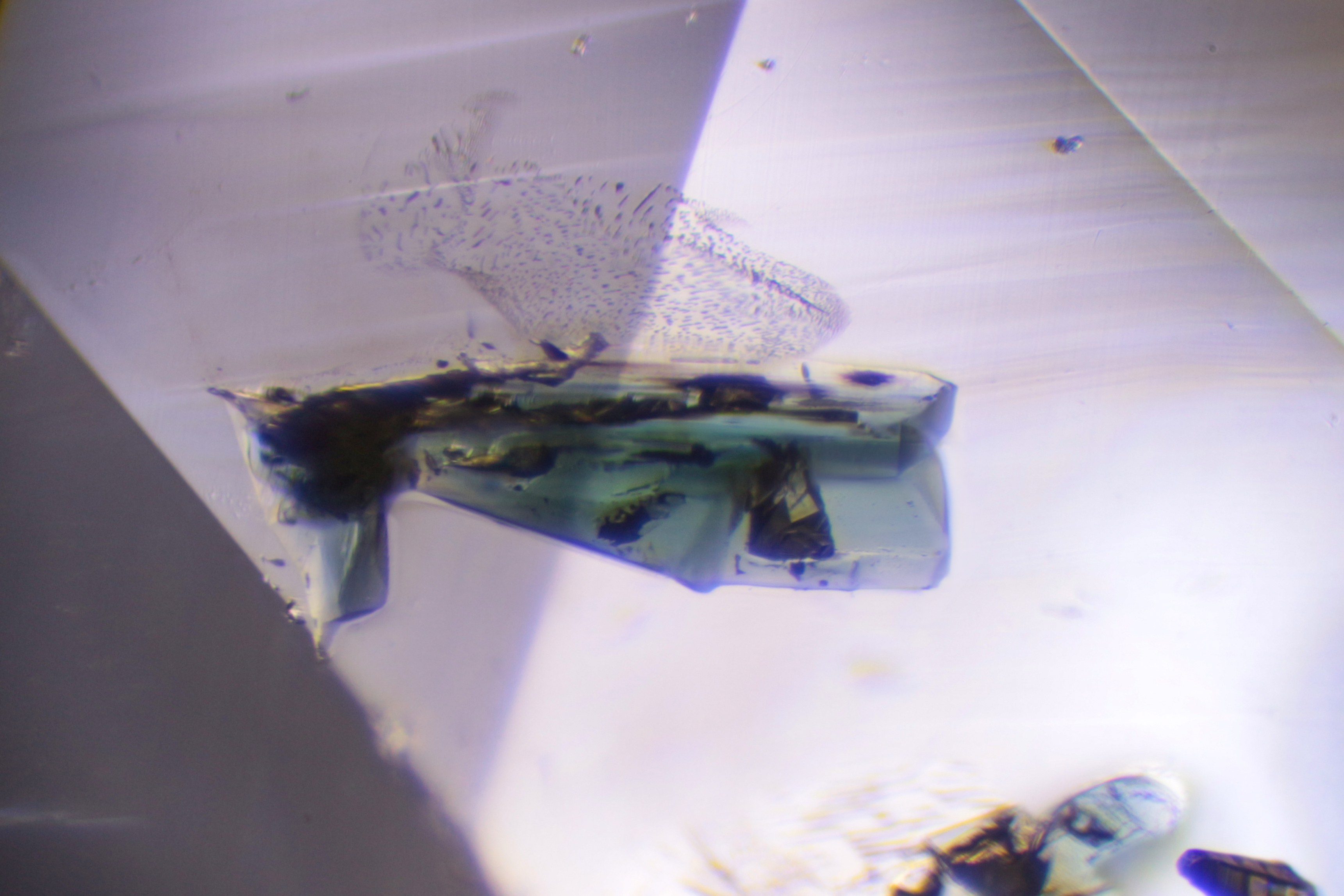
Microscopic examination revealed a diverse array of inclusions, which were identified by Raman spectroscopy as zircon, rutile, mica, graphite and amorphous carbon (e.g. Figure 41). Notably, none of these inclusions showed evidence of thermal alteration, indicating that the sapphire had not been subjected to high-temperature heat treatment.
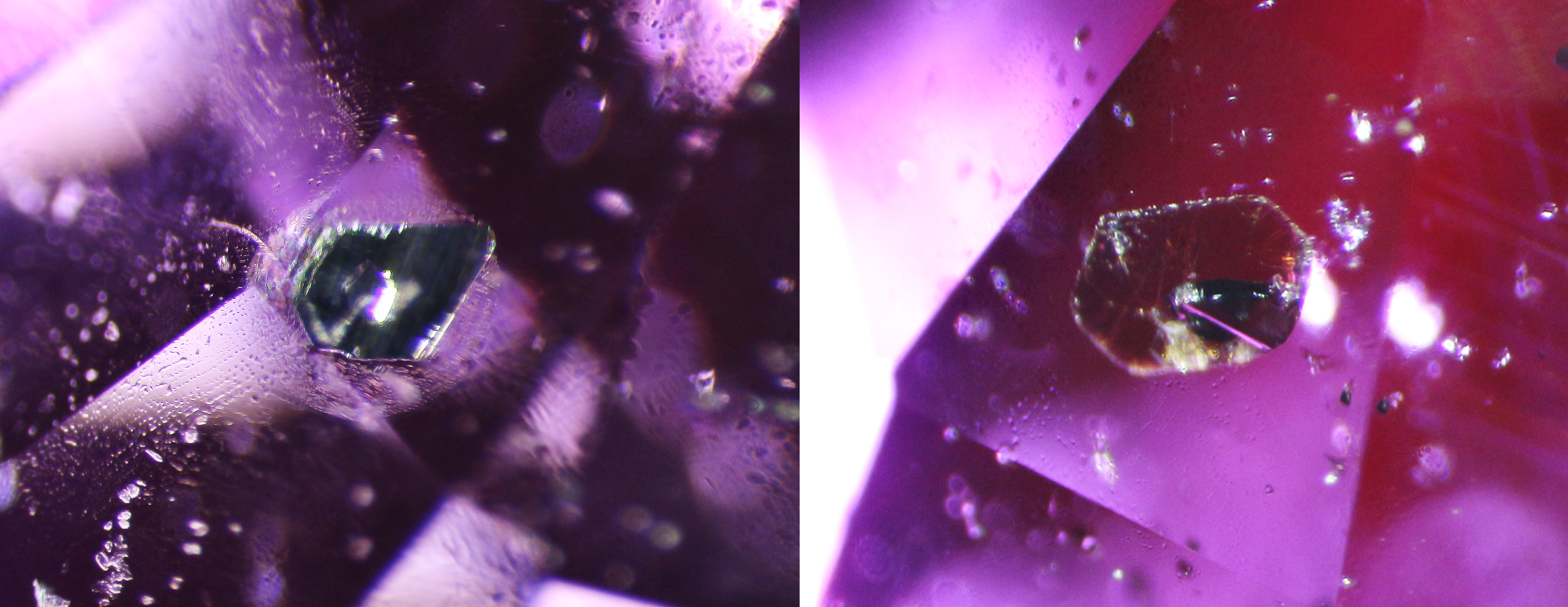
Figure 41: The sapphire contains a diverse array of inclusions, including (a) graphite and (b) mica, as well as zircon, rutile and amorphous carbon (not shown here). Photomicrographs by Fei Hu; image widths (a) 1.3 mm and (b) 1.4 mm.
A noticeable feature of this sapphire was a large eye-visible inclusion located just under the table, which was even more obvious under UV radiation due to its inert behaviour in contrast to the sapphire host (again, see Figure 40b). Initially thought to be a large negative crystal, further analysis using a Seamark X6600 Offline X-ray Inspection Machine indicated that the inclusion had a high density, as evidenced by its bright appearance in Figure 40c. This characteristic is consistent with inclusions such as zircon and rutile.
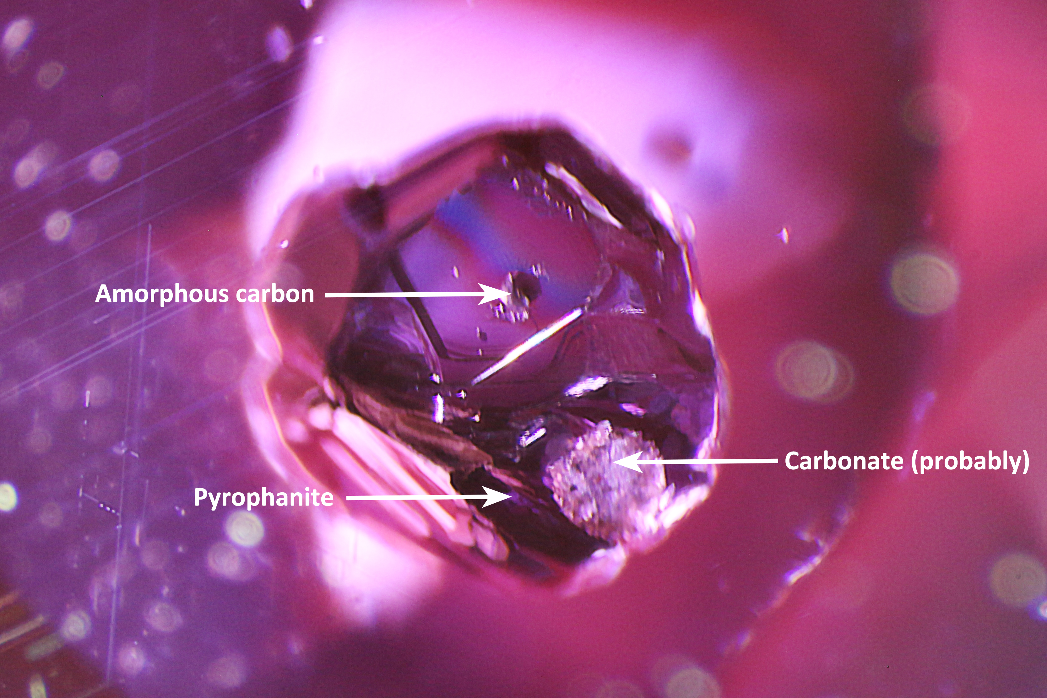
The most striking inclusion, however, was a relatively large, metallic-appearing crystal with well-developed morphology (Figure 42), setting it apart from the other inclusions. Raman spectroscopy could not provide a conclusive identification, possibly due to its depth within the host sapphire or strong scattering of the laser from its metallic-appearing surface. Closer examination revealed some additional mineral inclusions on its surface. One black spot was identified by Raman spectroscopy as amorphous carbon, and a white grain—most likely a carbonate—was visible nearby. In addition, a small, narrow, lower-lustre area on one side of the crystal yielded sharp Raman peaks at 164, 201, 235, 265, 330, 360, 463 and 685 cm-1, consistent with pyrophanite in the RRUFF database (Figure 43).
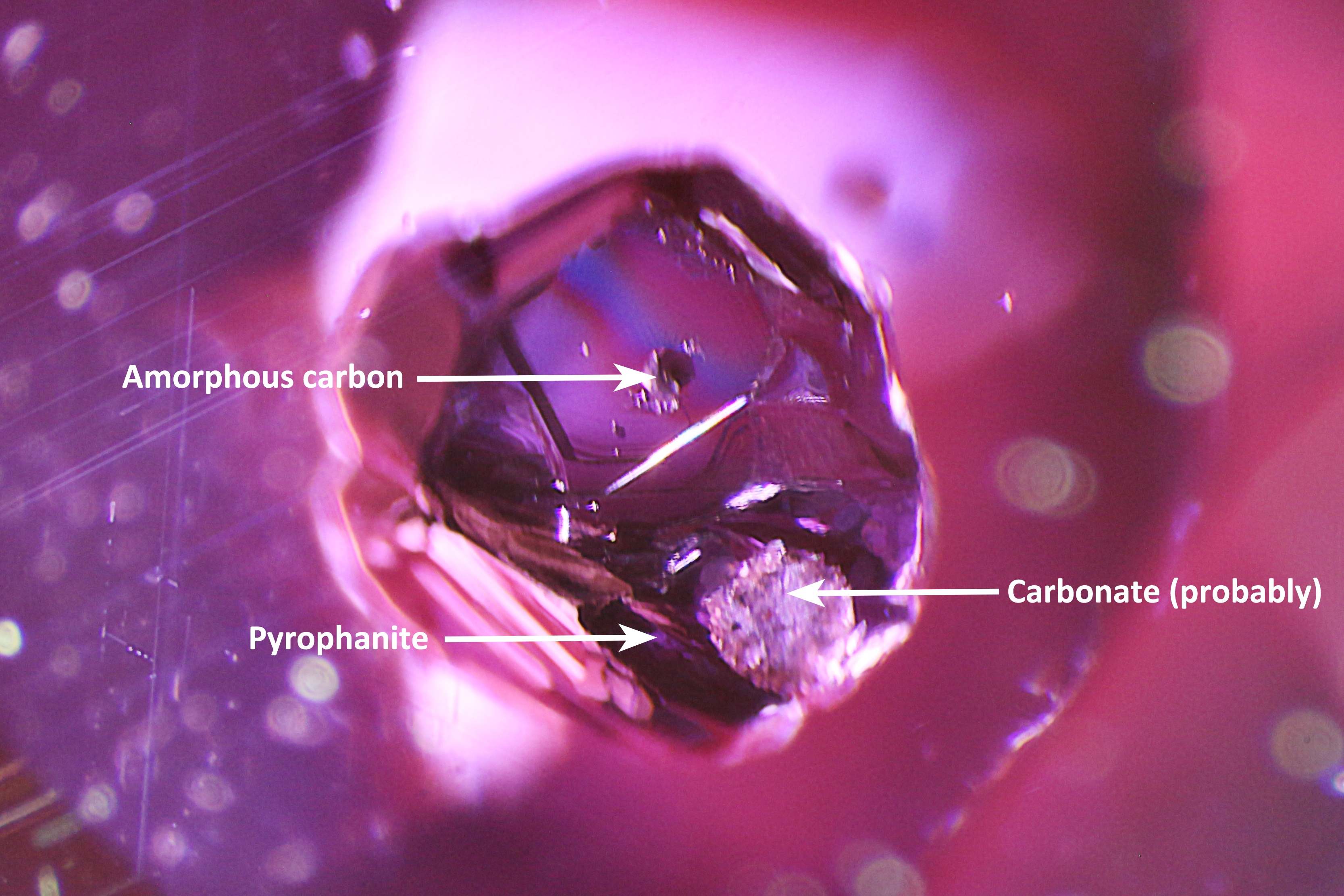
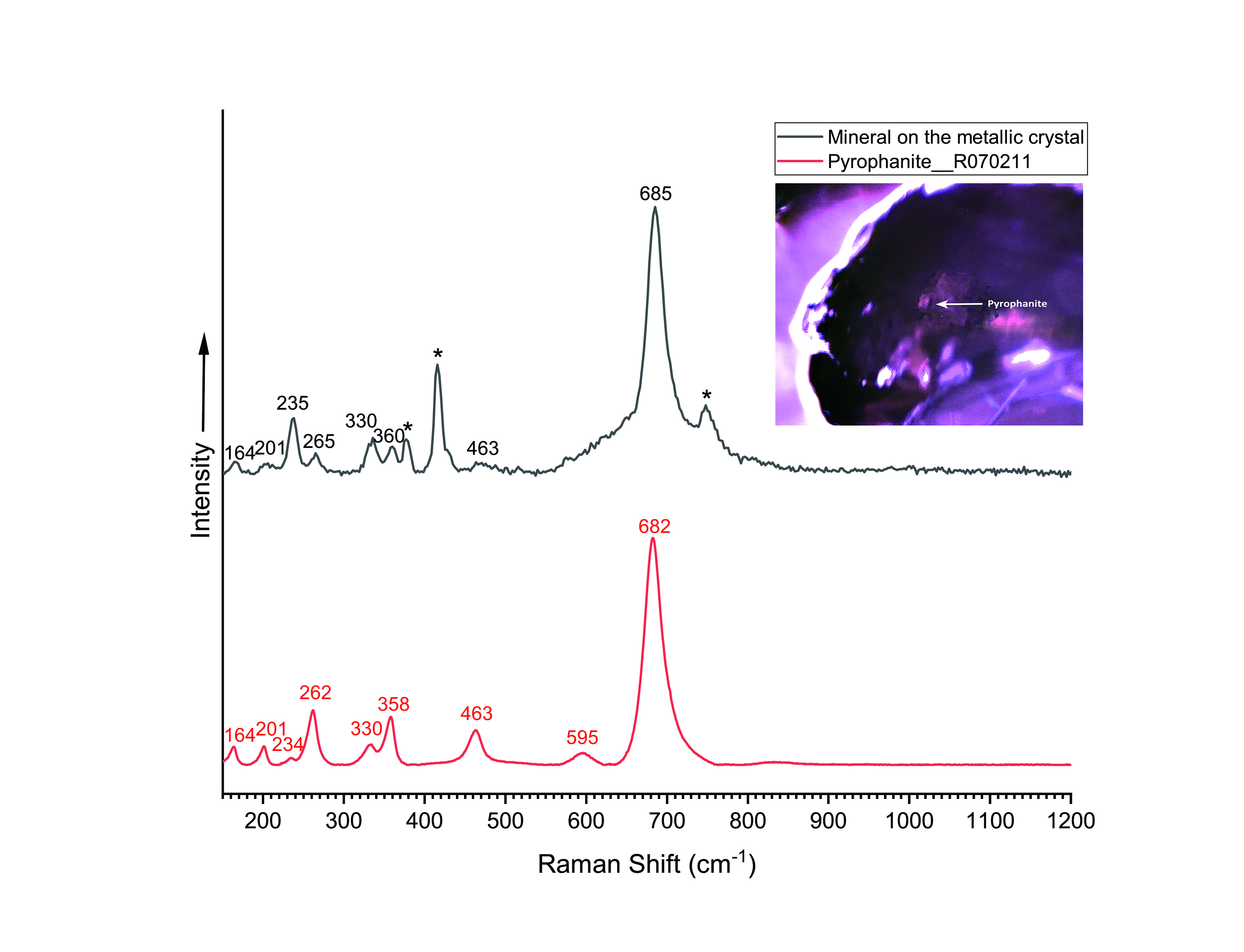
Pyrophanite is a rare manganese titanium oxide with the chemical formula MnTiO3 that forms a solid-solution with ilmenite (FeTiO3). It crystallises in the trigonal system and typically appears as reddish brown to dark red crystals. Pyrophanite forms in high-temperature metamorphic and hydrothermal environments, often coexisting with minerals such as ilmenite and garnet (Anthony et al. 2023). To the authors’ knowledge, this is the second documented occurrence—after Baldwin et al. (2017)—of pyrophanite as an inclusion in sapphire.
Geothermometry based on the Raman spectrum of a graphite inclusion in the sapphire suggests a formation temperature of around 850 ºC (cf. Huang et al. 2022), indicative of a relatively high-temperature environment. Furthermore, the sample exhibited a significant amount of Ti. These findings collectively suggest that this sapphire formed under reducing conditions in the presence of Ti, and at high temperature. The inclusions in this sapphire provide useful insights into the geochemical and environmental conditions governing its formation.
Yujie Gao and Ziyun Zhang
(zziyun@guildgemlab.com)
Guild Gem Laboratories
Shenzhen, China